
X-ray diffraction experiments are used to determine the structure of organic, inorganic and organometallic molecules.
We learn how the atoms in a molecule are connected to one another, as well as the three-dimensional arrangement of the molecules as they pack together to form as solid.
Further information about the facility is available here: Nanoscale Materials Characterization Facility | University of Virginia School of Engineering and Applied Science
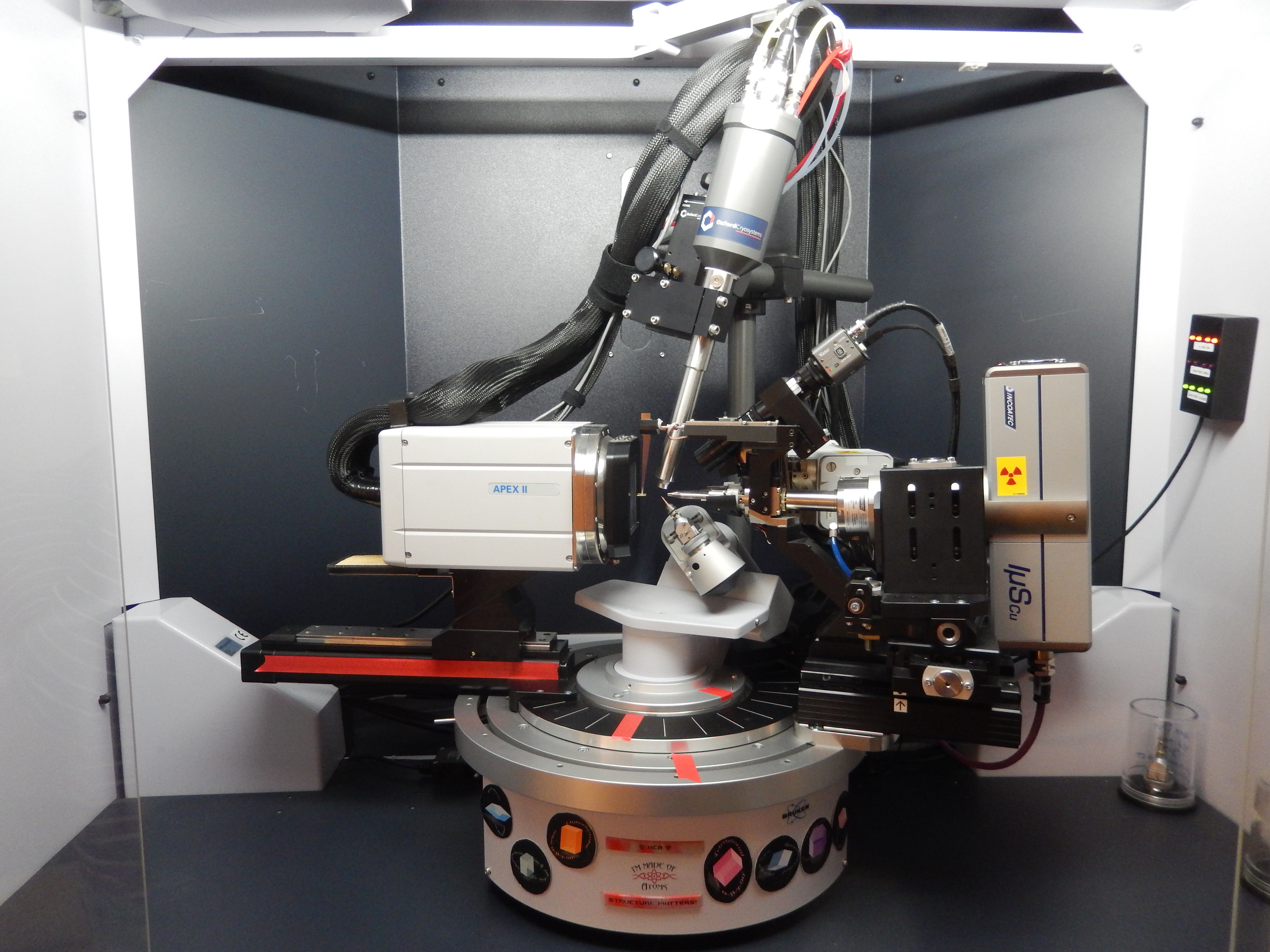
The department has one single-crystal dual-source X-ray diffractometer for the structural analysis of small molecules. Polycrystalline (powder) diffraction experiments and protein crystallography can also be done on this diffractometer. Three other diffractometers for polycrystalline samples (powders, thin films and/or bulk materials) are available through the Nanoscale Materials Characterization Facility.
Instrument Details:
Bruker ApexII Duo with Mo sealed tube (0.71 Å) and Cu IµS (1.54 Å) sources
Instrument Location:
Materials Science Building (MSE) room 108
Crystallographer Location:
Materials Science Building (MSE) room 103
Sample Submission Procedure:
All crystals must be accompanied by a sample submission form. Because it is impossible to predict in advance exactly how long it will take to collect a full dataset for any particular crystal, this instrument operates on a first-come, first served basis. To add your name to the waiting list, contact Diane Dickie. When the measurement begins on the crystal ahead of yours, you will be notified by email of the expected finishing time of that experiment, which becomes the start time for your experiment. Exceptions to the order of samples on the waiting list are rare, but could include postponing samples that are expected to need unusually long exposure times until the end of the day so that they can run overnight, or scheduling experiments related to classwork at the time that the class is held. Samples that are CLEARLY LABELED and stable at room temperature may be dropped off at any time. Samples that must be kept cold and samples that are best stored under an inert atmosphere should be kept in your research lab until it is time for your data collection. Please do not leave your samples in the X-ray lab after the experiment is finished. Remember, crystals don’t like vacuums and are often most stable when they’ve got a little bit of supernatant with them, so be careful about isolating and drying your sample.
Crystals are accepted from researchers outside of the UVA Chemistry department. Please contact Diane Dickie for current fees and shipping information. We offer unit cell screening, full data collection, and structure solution/refinement.
Training
Students whose research depends heavily on X-ray crystallography are encouraged to learn to collect, solve and refine their own structures. Structure solution and refinement is done with the SHELX software suite within OLEX2. Before you may operate the X-ray diffractometer, you must complete UVA’s Radiation Safety Training for the Safe Use of Analytical X-ray Equipment. You must also be up-to-date on your Chemical Safety and Waste Training. You will then get hands-on training with the diffractometer with supervision continuing until you have proven that you can safely operate the instrument and set up proper data collections independently. To get a more thorough background in X-ray crystallography, graduates students and upper-level undergraduates should consider taking CHEM5380, or attending a workshop like the American Crystallographic Association Summer School or the Canadian Chemical Crystallography Workshop.
FAQs
Q: How big should my crystal be?
A: Ideally between 0.1-0.5 mm in length, width and depth. Larger crystals can usually be cut to an appropriate size. Smaller crystals can often be successful, but may require longer data collection times.
Q: How much material do you need?
A: Only one good crystal is needed for the data collection, but it is a lot easier to find that one crystal if there are several to choose from. Try to bring at least a few mg if you can. It is possible to recover your leftover sample from the microscope slide by washing off the oil with an appropriate solvent if you have a very limited amount of material, but this may not be practical for compounds that are extremely air- and/or moisture-sensitive.
Q: How is the experiment done?
A: The submitted material is placed in a drop of oil and examined under a microscope. If an appropriate crystal is found, it is mounted on a reusable support (MiTeGen MicroLoop) and placed on the goniometer head under a stream of cold (100 K) nitrogen gas. After centering, a few frames of data are collected to judge the crystallinity and diffraction ability of the sample. Then a set of frames is collected to get enough data to determine the unit cell. The unit cell will be screened against the Cambridge Structural Database and any unpublished structures that are listed on the sample submission form. If no matches are found, a suitable data collection strategy is calculated and run. At the end of the collection, the data is integrated and scaled. The structure is then solved and refined.
Q: How long does it take to get results?
A: A typical data collection takes a few hours, but it could be as short as 20 minutes or as long as several days. Data processing and structure solution/refinement typically takes minutes to hours. It is almost always possible to get a preliminary structure solution before the data collection is complete, though, so you shouldn’t have to wait too long to get at least a rough idea of what your structure looks like.
Q: What do I get when my structure is completed?
A: You will get a CIF file, which is the file that you will eventually submit to a journal or database. You will get a report, which describes the details of the data collection, data processing, structure solution and structure refinement. It also contains tables of bond lengths, angles, and crystallographic information. You will get a checkcif file, which some journals require in addition to the CIF and which has information about the quality of the structure. You will get help preparing the structure for publication when you’re ready to do that. You will NOT get a picture of the structure unless you ask for it. Most people prefer to make their own pictures so that they can highlight what is important to them. All final results are sent to the student who submitted the crystal AND to the PI listed on the sample submission form.
Q: What information needs to be included on the sample submission form?
A: The sample label should match whatever is written on the sample container, so that it’s clear which form goes with which crystal. The sensitivity/stability information is needed so that the sample is handled appropriately. The other characterization section helps indicate how likely it is that that crystal really is what you think it is. The molecular formula is needed as a starting point for the structure solution. The color is helpful in case the sample changes between the time you submit it and the time it is run. The melting point, if known, will be included in the CIF file. The solvents are very important because solvent often gets trapped in the crystal lattice and could be a part of your final structure. The related structures are requested so that their unit cells can be compared with that of the current crystal to avoid collecting known compounds. The reaction pathway and proposed product are necessary because sometimes the thing that crystallizes isn’t what you expect but is a by-product or a starting material from earlier in the synthesis.
Q: How do I access the crystallographic databases?
A: There is a campus-wide web-access license through the library for both the Cambridge Structural Database (CSD) and the Inorganic Crystal Structure Database. You can also install the CSD on your own computer if you request the installation codes from either Diane Dickie (dad8v) or Jeremy Garritano (jg9jh). The data-processing computer in the X-ray diffraction lab has a copy of the PDF4+ database.
Q: How do I access other crystallographic software?
A: We have a department-wide (not campus-wide!) license for Diamond. You can download the software here and then contact Diane Dickie for a copy of the license. It runs on Windows only. You can download a basic version of Mercury as a stand-alone program here, or get the fully licensed version along with Conquest and other CCDC programs here after requesting the installation codes from either Diane Dickie (dad8v) or Jeremy Garritano (jg9jh). Most other software that we use is free for academic use and can be accessed after registering with each company. If you are planning to solve your own structures, you will need OLEX2, SHELX and Platon. Programs that may be of interest to highly experienced users include in Jana2006 and Max3D.
Q: How and when should I deposit my crystal structure in a database?
A: There are two options for depositing structures, and both are done here. Structures that will be included in a paper are deposited as pre-publication structures. This gets you a deposition number that can be included in the paper, but the structures are kept confidential until the paper is published. If more than one year goes by and your paper hasn’t been published, the database may contact you to make sure you’re still working on the paper. You can extend the confidential embargo as long as necessary. Structures can also be deposited as CSD Communications or ICSD Communications. Structures published this way are immediately publicly available in the database. They receive a permanent digitial object identifier (doi) and can be cited just like a paper, but they don’t require any additional writing. This route is recommended for structures that aren’t likely to be included in a traditional paper (starting material, by-product, crystal found in an NMR tube that was sitting in the back of a fume-hood for two years, etc.) and for structures that would otherwise be a very minor component in a paper focused on other, non-structural features.
Q: How do I publish my crystal structure?
A: Each journal has different guidelines for what is required when including a crystal structure in a paper. Some journals want you to deposit your CIF in a database such as the CSD or ICSD and then list the deposition number in the paper, while others ask for the CIF as part of your supporting information package. Some want each structure in its own CIF file, some ask for multiple structures to be combined into a single CIF. If your structure is “compound 3” in the paper, your future readers will thank you if you also publish your CIF as “compound 3” instead of the code it was assigned in the X-ray lab, especially when there is more than one structure in the paper. Requirements for figures showing crystal structures and tables of crystallographic data also vary from journal to journal. Check the author instructions as you’re writing your manuscript, and ask your crystallographer for help if needed.
Q: Should I include my crystallographer as a co-author on my paper?
A: Usually, yes because your crystallographer is interpreting data, not just collecting it. To get the refined crystal structure in your CIF, the crystallographer designed a proper data collection strategy, processed the diffraction images into useable data that accounts for the symmetry, absorption properties and imperfections (twinning, etc.) of your crystal, and used that data to determine the exact positions of every atom in your structure, including any that are disordered. This requires time and expertise, and meets the authorship definition for most scientific journals. If you do not feel that the crystal structure is a big enough part of your paper to merit co-authorship for your crystallographer, you should consider giving your crystallographer credit for the work by depositing the crystal structure in the appropriate database as a CSD Communication or ICSD Communication, and then citing that Communication in your paper. Remember to give your crystallographer enough time to review any manuscript drafts.